More than a century ago, a 26-year-old Albert Einstein explained Brownian motion in one of four papers he published in his annus mirabilis, the miraculous year, called because these papers shot him to fame. Brownian motion is the random jittering of small particles in a fluid, caused because they’re constantly colliding with molecules around them.
Now, scientists at the California Institute of Technology (Caltech) have developed a breakthrough imaging technique that enables real-time filming of these molecular motions. Their findings were published in Nature Communications.
‘Surreal experience’
Conventional microscopes are invasive and have limited fields of view. Other microscopes still can’t distinguish individual molecules, which are around tens of angstroms in size (1 angstrom = 0.0000000001 m). To compare, one human hair is about a million angstrom thick.
The Caltech team has now found a way to indirectly detect molecules by observing their interactions with light. Their technique also taps into the Brownian motion of particles.
Using the device they have reported that they can see down to tens of angstroms. “It was a surreal experience to visualise molecular sizes in real-time at the angstrom scale,” Yogeshwar Nath Mishra, who co-led the study when at Caltech’s Jet Propulsion Laboratory and who is now an assistant professor at IIT-Jodhpur, said.
“Even more remarkable was the realisation that no existing technique can achieve this level of detail.”
Need for speed
The more massive a particle, the slower its Brownian motion. “[It] is like watching how much a spinning object twists after being nudged by light. Small molecules spin fast and scramble the light more. Big molecules spin slowly and keep it aligned,” Lihong Wang, director of the Caltech Optical Imaging Laboratory and who supervised the study, said.
So by measuring how fast a molecule changes the properties of light, they could determine its size.
The Egyptian-American chemist Ahmed Zewail from Caltech was the first to measure particle motion at super-short time scales. This work allowed his team to observe chemical reactions as they happened for the first time. He was awarded the Nobel Prize for chemistry in 1999.
“While traditional techniques often rely on time-consuming point-by-point scanning, our approach captures the scene in a single shot,” Wang said. “We also achieved imaging speeds of hundreds of billions of frames per second, making it possible to observe molecular interactions in unprecedented slow motion.”
The device is thus the world’s fastest single-shot microscope.
“Finally, unlike [traditional methods] which require extensive sample preparation and often damage the specimen, our method is non-intrusive, enabling direct, in-situ measurements,” Wang added.
“Some of the most exciting features of this microscope include its wide-field imaging capability, offering an image area of a few square centimetres, an order of magnitude larger than conventional microscopes,” per Mishra. “To the best of our knowledge, our work is the first ever to achieve the feat of single-shot 2D molecular sizing.”
Playing jigsaw
They tested their microscope using a molecule called fluorescein-dextran. Fluorescein is a food colouring dye. Fluorescein-dextran is used to monitor blood flow, drug delivery, and tissue and cell labelling. These fluorescent molecules come in the form of powders. The scientists blended them with water and used clean pipettes to pour drops of these samples into cuvettes (clear, short, rectangular tubes for holding liquid samples).
Florescein powder dropped into a solution of tapwater glows a bright green under a typical blacklight after around 15 seconds.
| Photo Credit:
Bricksnite (CC BY)
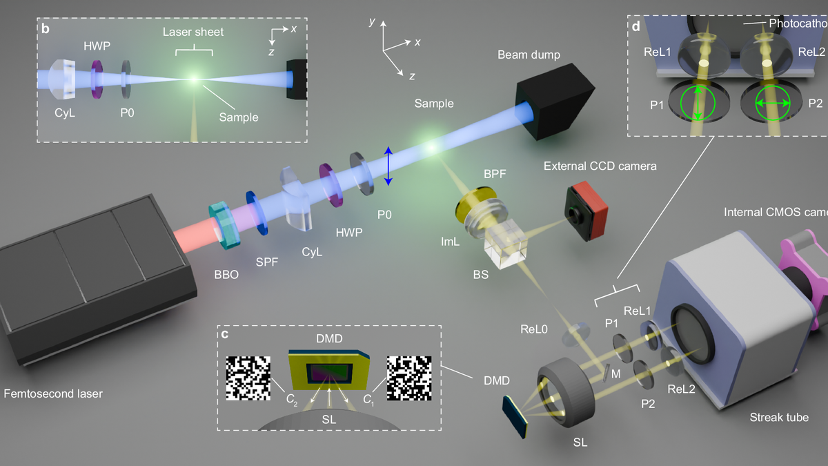
Then they turned to ultrashort pulses from a laser. These lasers aren’t unlike those used in LASIK and cataract surgeries. The laser sheet slices through the sample in the cuvette. As it does, the sample emits light that falls on an array of small square mirrors making up a digital micromirror device (DMD).
The DMD’s job is to shape the light beam. Researchers use software code to tilt each individual mirror in this light-crafter depending on the corresponding pixel in the input image.
“Imagine you’re trying to solve a jigsaw puzzle, but instead of having all the pieces, you only have a few of them — and surprisingly, you can still figure out what the full picture looks like,” Wang said.
This idea underpins the team’s technique, which can reconstruct the full picture from very few measurements provided the structure is repetitive. The DMD converts the transient scene into a random jigsaw pattern from which researchers can extract information about the full picture.
The light finally passes through a streak tube that converts the photons in light to electrons. A phosphor screen collects these electrons as they sweep across it and creates a pattern of streaks. The streak pattern reveals the pulse duration from which scientists can infer the sizes of the molecules.
Ensemble of molecules
“It is an interesting piece of work. The key in this work is the use of the streak camera to detect dynamics in nanoseconds. This is within the actual lifetimes of the molecules and wouldn’t be possible with slow detectors or photodetectors,” Basudev Roy, an associate professor at IIT Madras who works on super-resolution microscopy and wasn’t involved in the recent study, said.
The size of molecules measured using their technique concurred with previous estimates. “It still sees an ensemble of molecules inside a detection region — it still doesn’t see a single molecule yet. But the dynamics indicate chemical compositions and also chemical reactions,” Roy said.
“Surprisingly, we found out that the technique also works in gas phases. … Initially, we assumed it would be challenging to apply [it] in turbulent environments, such as within a flame,” said study co-lead Peng Wang of Caltech.
The team observed black carbon nanoparticles in flames through the microscope. “Our data in the gas phase turned out to work excellently and the molecule size matches … experimental observation well,” Peng said.
This new imaging technique could help better visualise processes and transform biomedical research, disease detection, drug design, and nanomaterial fabrication, among others.
Unnati Ashar is a freelance science journalist.
Published – July 28, 2025 05:30 am IST