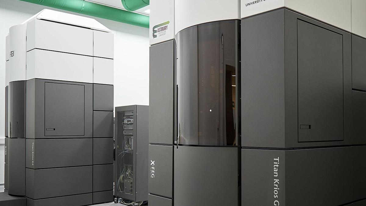
A pair of cryo-electron microscopes at the University of Leeds.
| Photo Credit: Hiramano92 (CC BY-SA)
Scientists use a powerful technique called cryo-electron microscopy to see the 3D shapes of biological molecules, but it normally needs the molecules to be extremely concentrated in a sample first. But for rare molecules this is hard to achieve.
In a new study, researchers in the U.S. have created a workaround called Magnetic Isolation and Concentration cryo-electron microscopy (MagIC for short). It lets researchers sidestep the limitation and study samples 100x more dilute than before. The findings were published in eLife in May.
The new method works by attaching molecules of interest in a sample to 50-nm beads, then using a magnet to clump the beads together. This way each micrograph ended up with several usable images even when the solution had less than 0.0005 mg/ml of the molecules.
Because the beads were easy to spot even at low magnification, the scientists could quickly move the microscope to regions rich in particles, speeding up data collection.
Small particles often hide in background noise. To pull them out, the authors built a computer workflow called Duplicated Selection To Exclude Rubbish (DuSTER). It picked each particle twice, kept those that landed in the same place after two rounds of 2D or 3D classification, and threw the rest away.
Thus MagIC lowers the sample demand to just 5 nanograms per grid while DuSTER rescues clear classes from seemingly hopeless images.
Published – June 09, 2025 05:45 am IST
