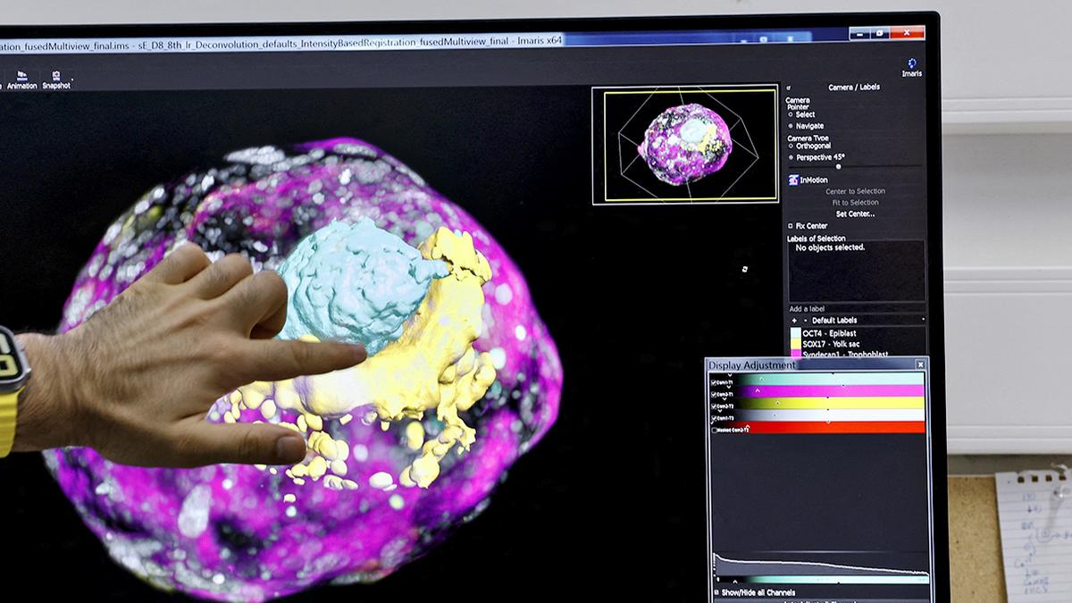
Image used for representation
| Photo Credit: Amir Cohen
(This article forms a part of the Science for All newsletter that takes the jargon out of science and puts the fun in! Subscribe now!)
Embryo implantation is a decisive step in mammalian reproduction. When a fertilised egg reaches the uterus, it must successfully attach to and invade the maternal tissue for pregnancy to continue. However, implantation often fails: an estimated 60% of miscarriages are due to problems at this stage. Traditionally, scientists have investigated implantation by looking at genes and chemical signals that control how embryonic cells behave. While valuable, this focus has had a gap: implantation is also at its core a physical act. An embryo must push, pull, and burrow into the tissue that surrounds it.
Human embryos also invade deeply into the uterine wall, embedding themselves almost entirely. In contrast, mouse embryos attach more superficially, settling into crypt-like spaces rather than tunneling all the way in. These differences affect placental development and pregnancy outcomes. Understanding why they exist and how mechanical forces shape them can illuminate human fertility challenges.
Studying these forces directly in living embryos has been nearly impossible, however. Implantation occurs inside the uterus, hidden from view, and the tools to measure feeble forces in such delicate systems have been lacking. A team of scientists from the Institute for Bioengineering of Catalonia in Spain recently reported in Science Advances a solution to this problem.
The researchers designed an artificial environment outside the body that mimics the uterine lining. They built a flat, 2D collagen gel and a 3D collagen matrix, both resembling the fibrous extracellular tissue embryos naturally encounter. Human and mouse embryos were placed on or within these gels. Then, advanced imaging tools captured how the embryos pulled, pushed, and deformed the surrounding material.
Computational methods tracked small shifts in the fibers, producing colour-coded maps of how the forces spread. The setup allowed researchers to see embryos developing and measure the traction they exerted on their environment in real time.
The experiments revealed that embryos don’t passively sit in the uterus: they actively reshape it. Both mouse and human embryos generated pulling forces that reorganised collagen fibers around them. While mouse embryos produced strong, directional pulls along two or three main axes, human embryos embedded deeply into the matrix, creating multiple small focal points of traction that spread radially. In other words, mice pulled outward and humans pulled inward.
Low-quality human embryos (those that were smaller or contained dead cells) produced weaker forces and failed to invade properly, suggesting force generation is a marker of healthy development. Additional tests showed that disrupting the proteins that connect embryonic cells to the matrix reduced force transmission, confirming that these attachments are critical. When scientists pressed on the gel with a needle, human embryos also sent protrusions towards the pressure point.
The findings indicate that mechanical forces are not side effects but drivers of early development. Clinically, the findings open new directions for fertility research. If healthy implantation depends on embryos generating certain patterns of force, doctors could one day use mechanical signatures to assess embryo quality during in-vitro fertilisation. Such tools could also improve success rates while reducing the emotional and financial toll of repeated treatments.
From the Science pages
Question Corner
Flora and fauna
Published – August 20, 2025 12:31 pm IST
